

Molecular Services and Biofilm Research
We specialize in analyzing medical biofilms using FISH (fluorescence in situ hybridization) and nucleic acid-based methods such as PCR, sequencing, and microbiome analyses. Combining visualization with genomic and microbiome data provides entirely new insights into infections and biofilms.
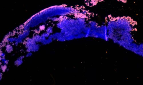
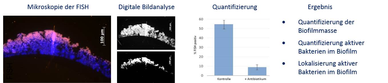
FISH is a molecular biology method that allows you to visualize and measure the effectiveness of antimicrobial substances on biofilms. Without relying on culture methods, it quantifies the effect with spatial precision: Where does the substance act? How does it work? Are any microbes surviving? And how does the killing process depend on time and dosage within the biofilm?
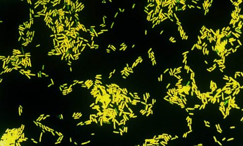
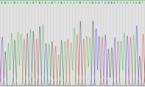
We provide identification of cultures and microorganisms in tissue samples using pan-bacterial 16S rRNA gene amplification followed by Sanger sequencing. In this process, specific regions of the bacterial DNA, such as segments of the 16S rRNA gene, are amplified through PCR (Polymerase Chain Reaction). Sequencing these amplified regions then allows precise identification of the microorganisms.
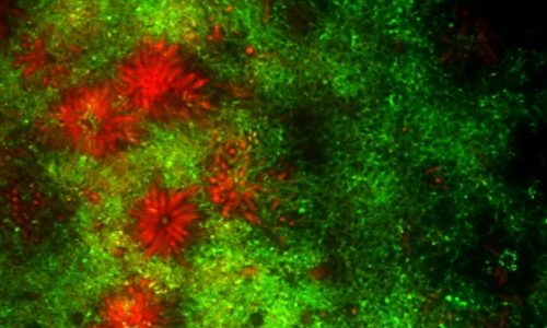
In mixed biofilms, we combine FISH with microbiome analyses to study all microorganisms in a sample. DNA from methacrylate-embedded samples can be amplified with high sensitivity and minimal risk of contamination. Using FISH results together with sequencing data greatly improves the precision of microbial identification.